
Proper clinical documentation is the most efficient way to avoid revenue loss for radiologists. Ensuring proper documentation also helps recoupment in the event of an audit, and prevention of claims denial due to insufficient documentation. This series of tips dissects common documentation errors in the radiology setting, which include but are not limited to failure to enumerate body parts, missing documentation for ultrasound (US), lack of documentation of 3-dimensional (3D) reconstructions for computerized tomography (CT), CT angiography (CTA), magnetic resonance imaging (MRI) and magnetic resonance angiography (MRA), missing spectral analysis and color flow information for duplex Doppler, failure to document permanent images for US guidance, and use of equivocal language and vague clinical indications. Here we focus on the documentation needed for duplex Doppler examinations, using examples that are coded to Current Procedural Terminology (CPT).
According to the American Medical Association (AMA), which publishes the CPT, a duplex Doppler US is performed for characterizing the pattern and direction of blood flow with the production of real-time images integrating 8-mode 2D vascular structure with spectral and color flow Doppler mapping or imaging [Clinical Examples in Radiology, Summer 2012]. 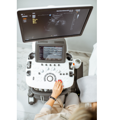
Documentation of spectral analysis, color flow and grayscale must exist to support a duplex doppler. You may see the following terms, for example:
- Imaging: B-mode; diagnostic evaluation; real-time imaging or 2D imaging
- Spectral analysis: Acceleration rate; bandwidth broadening; mono, bi or tri; peak systolic velocity; phasicity; resistive index (RI); spectral broadening; velocity; waveform analysis; pulsed Doppler or pulsatility
Missing documentation of any one of the three required components will cause the claim to go out as a plain US, e.g., CPT code 76882 [Ultrasound, limited, joint or other nonvascular extremity structure(s) (e.g., joint space, peri-articular tendon[s], muscle[s], nerve[s], other soft-tissue structure[s], or soft-tissue mass[es]), real-time with image documentation], which is reimbursed at about 70% of CPT code 93970 [Duplex scan of extremity veins including responses to compression and other maneuvers; complete bilateral study].
Again, you can see the importance of complete documentation towards avoiding revenue loss for radiologists.